
Les 25 meilleures idées de la catégorie Anatomy of the face sur
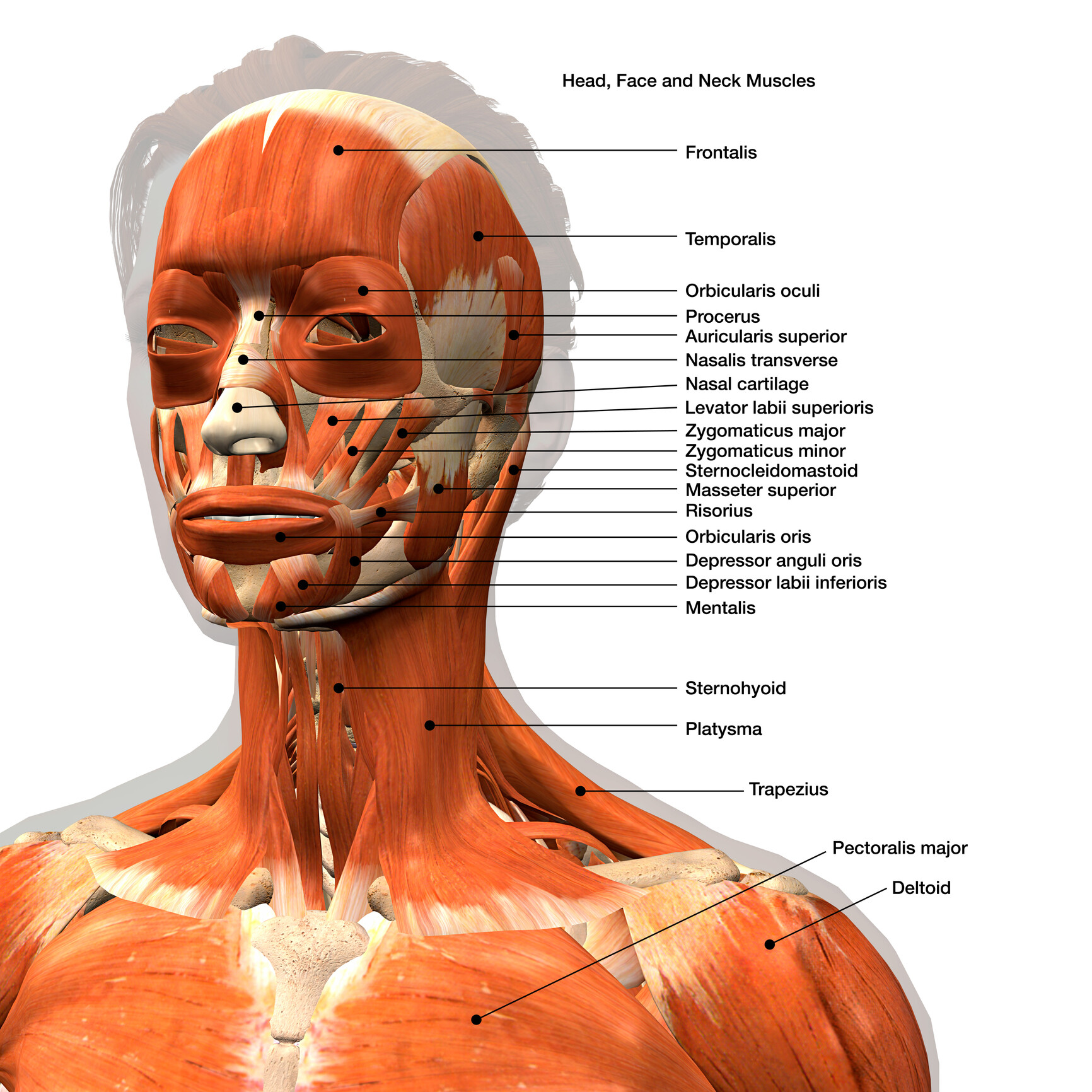
The facial muscles (also called the muscles of facial expression) are situated within the subcutaneous tissue of the face. They are responsible for the movements of skin folds, providing different facial expressions. The facial muscles originate from the bones of the facial skeleton (viscerocranium) and insert into the skin.. These muscles are mostly grouped around the natural orifices of the.

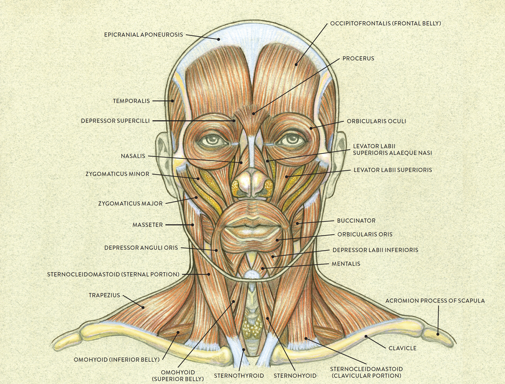
Anatomy of the facial muscles. Reprinted under Creative Commons
Facial anatomy at-a-glance Dec 06, 2017 Knowing the facial anatomy is fundamental to performing more than aesthetic surgery. A provider's lack of understanding of the intricate web of facial muscles, nerves, arteries and more can turn a relatively simple injection technique, with botulinum toxin or a filler, into a serious complication.

Medical Science on Instagram “Wow! 😱Look at the amazing detail of the
The curved part of a bone that gives structural support to the rest of the bone. Above: Markings of the facial bones with the following views: (A) anterior view, (B) lateral view of the left side of the skull, (C) inferior view with the mandible removed, and (D) lateral view of the right side of the skull. Marking.

Pin em Pilates
The facial muscles, also called craniofacial muscles, are a group of about 20 flat skeletal muscles lying underneath the skin of the face and scalp. Most of them originate from the bones or fibrous structures of the skull and radiate to insert on the skin.

FileLateral head anatomy.jpg Wikimedia Commons
The facial vein is a large vessel of the face and is much less tortuous than the artery of the same name. It lies posterior to the facial artery and begins from the lateral side of the nose. It drains the external palatine vein and will go on to join the retromandibular vein. This then forms the common facial vein.

Illustration Of The Anatomy Of A Female Human Face stock photo
Development of the Face and Palate. The external human face develops between the 4 th and 6 th week of embryonic development. The development of the face is completed by the 6 th week. Between the 6 th and 8 th week, the palate begins to develop. Consequently, this causes a distinction between the nasal and oral cavities.

Face anatomy by JosueVilela Medical Visualization Sculpture CGSociety
Structure and Function The anatomy of the face can divide into three main regions: upper face, middle face, and lower face. The entire face is covered by skin superficially, while the deep anatomy contains muscles, fat pads, nerves, vessels, and bones. Upper Face
Muscles Of Face Diagram
ISSN 2534-5079. We attempted to synthesize the anatomy of the face and neck in this anatomy module. We used MRI images T2-weighted with axial, sagittal and coronal planes. 512 anatomical structures were dynamically labeled, and some structures have been redesigned or enhanced with a graphic tablet for better readability.
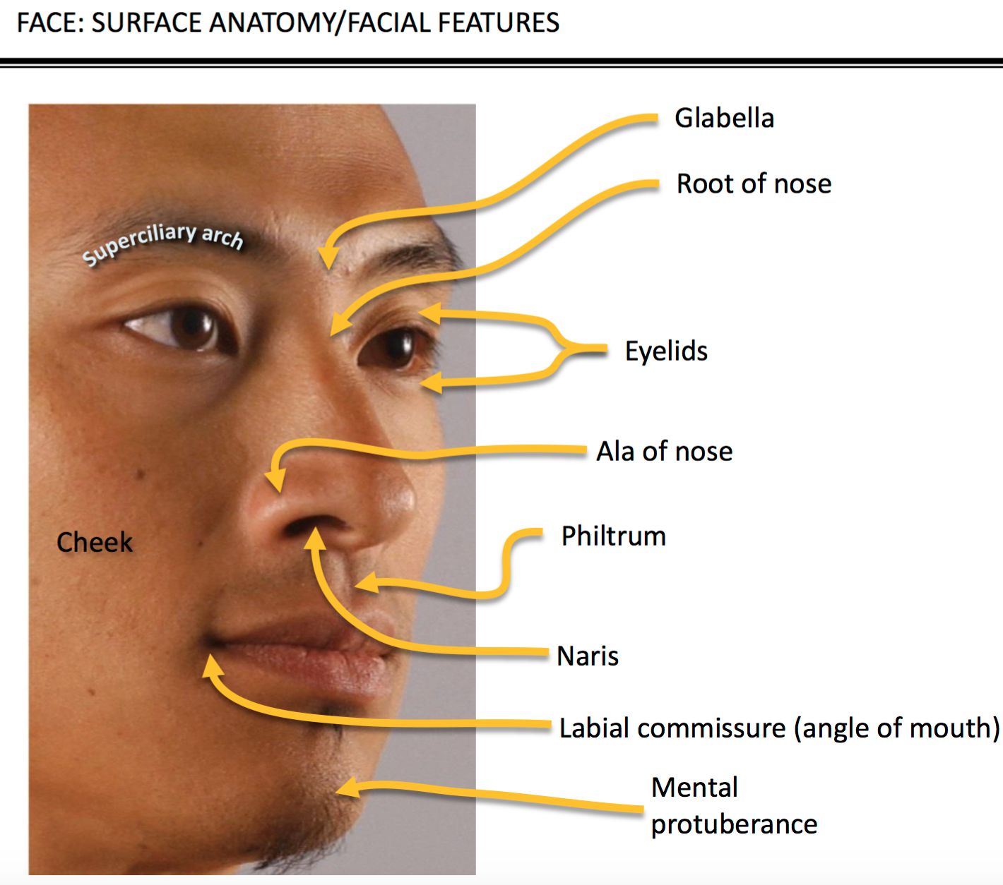
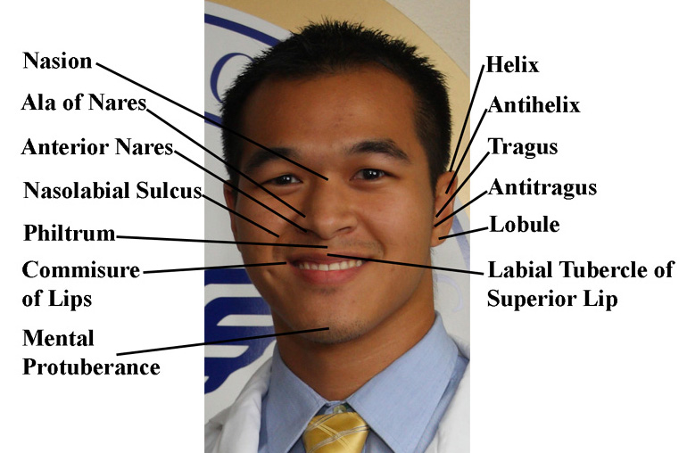
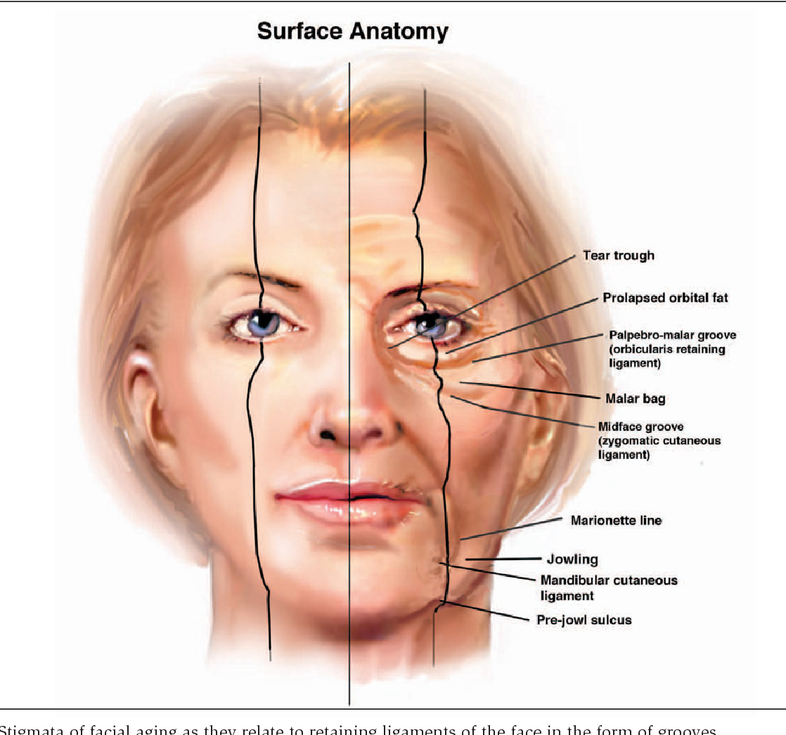
Surface Anatomy Of Face
I show you the muscles and bones of the human face. ︎Support me on Patreon: https://www.patreon.com/angelganev ︎ ︎Watch the FREE livestream: https://www.yout.

Resultado de imagen para face anatomy Facial Anatomy, Head Anatomy
F a c i a l B o n e s Functions Provide a structural framework for the face. So, the look or form of our face is due to our facial skeleton. Support the soft tissues of the face, head, and neck. Protect a part of the brain. Encase and protect the sensory organs — nose, eye, and tongue.
Discover the Hidden Secrets of Your Face Muscle Anatomy Drawing to Amp
Download the Temu App and start saving more today! Unleash incredible deals and coupons. Ready to shop and save? Explore amazing deals on the Temu App. Free shipping & return.
Second Language Diary of a Caribbean Med Student
Anatomy of the viscerocranium. Viscerocranium Synonyms: none The skull (cranium) is a complex bony structure composed of two distinct regions: the neurocranium and viscerocranium. The viscerocranium is a collection of bones that make up the face skeleton.

Anatomical Head Model, Anatomical Human Anatomical Half
Skeletal anatomy of the face The face is the feature which best distinguishes a person. Specialized regions of the human brain, such as the fusiform face area (FFA), enable facial recognition; when these are damaged, it may be impossible to recognize faces even of intimate family members.
MUSCLES OF THE FACE—LATERAL VIEW
In human face anatomy, all the features curve up and the ear moves up. Because the nose juts out, it oversteps its line (see figure) and the tip looks much closer to the mouth—if the face turns down enough, the nose will squarely overlap the mouth. Seen from this angle, the nose displays no details at all, just the wedge with a hint of wings.
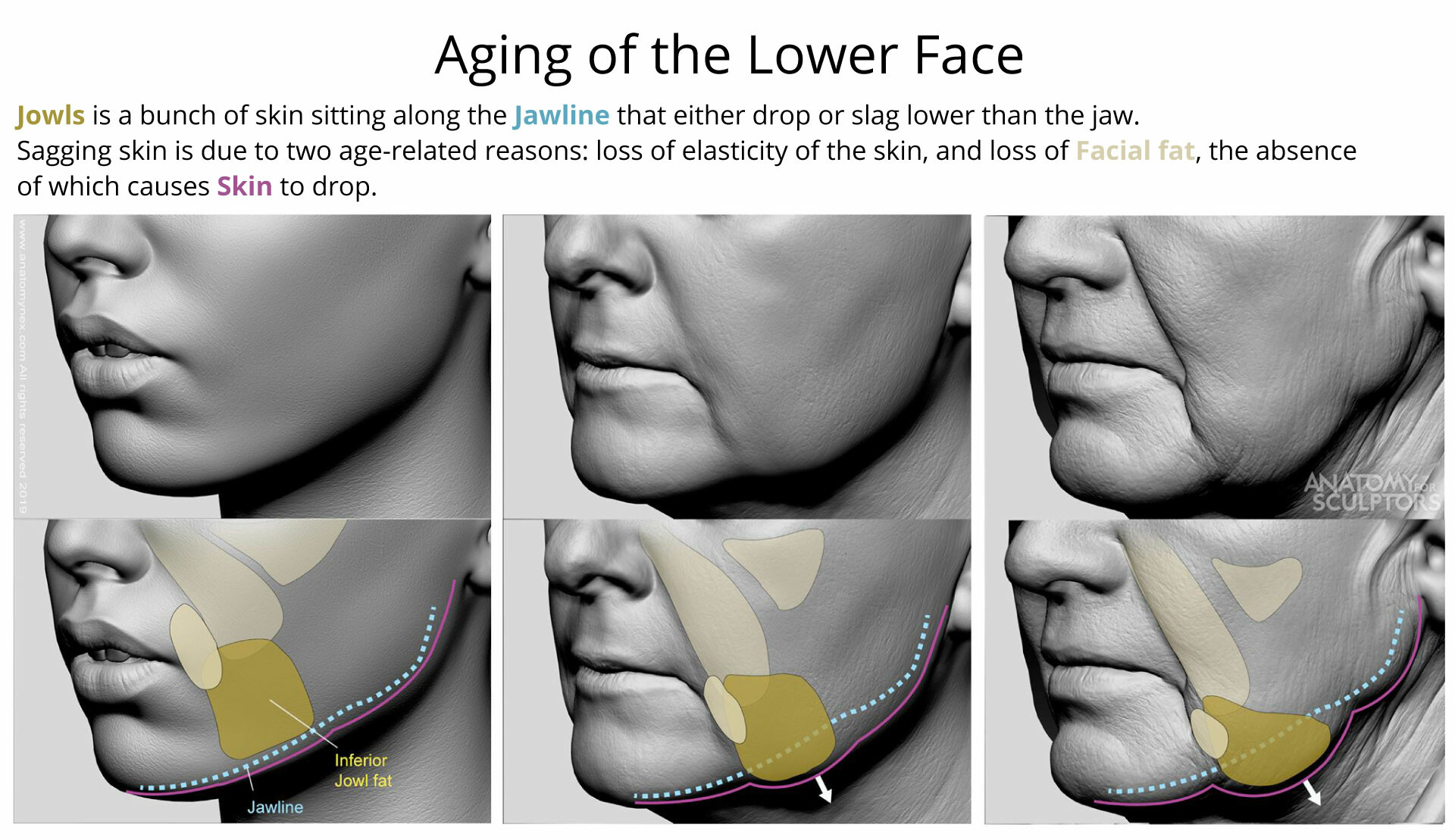
ArtStation Aging of the Lower Face
Face Anatomy Anatomy Head and Neck Areas Face Anatomy Contents Skin of the Face Clinical Significance of the Skin of the Face Cleavage Lines of Skin in the Face Superficial Fascia of the Face Deep Fascia of the Face "Face Characteristic Features of the Muscles of Facial Expression Location and Function Muscles Around the Orifice of the Eye
Anatomy Of Face Anatomical Charts & Posters
face, front part of the head that, in vertebrates, houses the sense organs of vision and smell as well as the mouth and jaws. In humans it extends from the forehead to the chin. During the course of evolution from the prehuman Australopithecus to modern humans ( Homo sapiens ), the face became smaller in relation to the overall size of the head.